Monthly Archives: October 2026

AbMole Mini-Lecture | Treosulfan (Treosulphan): A Highly Efficient DNA Crosslinking Agent and Its Applications in Cancer Research


Treosulfan (Treosulphan, NSC 39069, AbMole, M13635) is an alkylator that undergoes non-enzymatic hydrolysis under physiological conditions to generate active metabolites, exhibiting significant cell proliferation inhibitory effects. It serves as a critical tool in various tumor models and hematopoietic stem cell transplantation studies. The chemical structure of treosulfan features a six-carbon sugar alcohol backbone, in which four hydroxyl groups are methylated, the hydroxyl group at the C2 position is substituted with a sulfhydryl group, and sulfate ester groups are attached to the C1 and C4 positions, respectively. This structural design imparts chemical stability under neutral pH conditions, whereas hydrolysis occurs under weakly acidic environments: the sulfate ester groups are first removed to generate an intermediate monoepoxide derivative (EBDM), which subsequently undergoes spontaneous cyclization to form the electrophilic diepoxide metabolite (DEB). This activation process does not require enzymatic catalysis. As the ultimate active form of treosulfan (NSC 39069), DEB attacks nucleophilic sites (primarily the N7 position of guanine) in DNA molecules, forming intra- and interstrand crosslinks. These crosslinks distort the DNA double helix, impede the normal functions of DNA polymerases and topoisomerases, and ultimately induce S-phase cell cycle arrest and activation of apoptotic pathways. Compared to traditional nitrogen mustard alkylating agents, the diepoxide structure of treosulfan exhibits higher DNA crosslinking efficiency, with IC₅₀ values reaching micromolar or even nanomolar levels in various tumor cell lines.

Treosulfan (Treosulphan, AbMole, M13635) has garnered extensive attention in the field of hematopoietic stem cell transplantation. Treosulfan effectively eliminates host hematopoietic cells while causing minimal damage to non-hematopoietic tissues. For instance, in NOD/SCID mouse models, preconditioning with treosulfan (CAS No.: 299-75-2) (1.5 g/kg/day for 3 days) significantly improves the engraftment success rate of CD34⁺ cells. Beyond hematologic malignancy research, treosulfan (NSC 39069) is also applicable to cell- and animal-level studies of other tumors. For example, treosulfan in combination with fludarabine synergistically kills ovarian cancer cell lines SKOV3 and OVCAR4 [1]. In an Ewing’s sarcoma A673 cell xenograft model, intraperitoneal injection of treosulfan (150 mg/kg, twice weekly) reduces tumor volume by 64% while downregulating the expression of ETS fusion genes [2].


References and Acknowledgments

[1] I. El-Serafi, I. Micallef Nilsson, A. Moter, et al., Impact of fludarabine and treosulfan on ovarian tumor cells and mesothelin chimeric antigen receptor T cells, Cancer immunology, immunotherapy : CII 73(9) (2024) 163.

[2] Sebastian Werner, Arnulfo Mendoza, Ralf A Hilger, et al., Preclinical studies of treosulfan demonstrate potent activity in Ewing’s sarcoma, 62(1) (2008) 19-31.


AbMole Mini Lecture | Selisistat (EX527): A Core Tool Compound for Exploring SIRT1 Function


Selisistat (EX527, AbMole, M1708) is a potent and highly selective inhibitor of SIRT1 (Sirtuin 1). Selisistat (CAS No.: 49843-98-3) binds to the complex of SIRT1 and nicotinamide adenine dinucleotide (NAD+), thereby blocking the deacetylase activity of SIRT1. In research, Selisistat serves as a key tool for investigating SIRT1 function. For example, in metabolic studies, Selisistat (EX527) was used to reveal that SIRT1 regulates PPARα protein stability, showing that SIRT1 inhibition prevents ubiquitin-mediated degradation of PPARα[1]. In tumor models, Selisistat, alone or in combination with Paclitaxel, inhibits tumor growth via mechanisms involving cell cycle regulation and metabolic reprogramming[2]. In neuroscience, Selisistat (EX527) influences microglial phenotype switching by inhibiting SIRT1, thereby altering the production of inflammatory cytokines and reactive oxygen species[3]. In virology, downregulating SIRT1 activity with Selisistat significantly inhibits hepatitis B virus replication[4]. Selisistat also affects various other cellular processes through SIRT1 inhibition. For instance, in inflammatory responses, Selisistat (EX527) reverses the SIRT1-activating effects of Resveratrol, thereby attenuating its anti-inflammatory activity[5]. In oxidative stress models, Selisistat treatment reduces mitochondrial function and promotes apoptosis[6]. Selisistat has also been used to elucidate the role of SIRT1 in autophagy[7]. In skin hair follicle regeneration studies, Selisistat promotes hair regrowth by inhibiting the ferroptosis pathway [8].

Case Study

Int J Biol Sci. 2022 Jan 31;18(4):1594-1611

In the above study, researchers from the Shanghai Institute of Materia Medica, Chinese Academy of Sciences, and Shanghai University of Traditional Chinese Medicine investigated the potential of Atractylenolide III (ATL III), a bioactive compound derived from Atractylodes macrocephala, to inhibit non-alcoholic fatty liver disease (NAFLD) and its possible mechanisms. The study found that ATL III alleviated liver injury and lipid accumulation in high-fat diet (HFD)-induced NAFLD mouse models and free fatty acid (FFA)-treated HepG2 cells by activating the AdipoR1-mediated AMPK and SIRT1 signaling pathways. In this study, researchers used Dorsomorphin (Compound C, AbMole, M2238) and EX 527 (Selisistat, AbMole, M1708) as inhibitors of the AMPK and SIRT1 pathways, respectively, confirming that both pathways are essential for the biological activity of Atractylenolide III. In cell experiments, the CCK-8 kit (AbMole, M4839) was also used to verify the effect of Atractylenolide III on the viability of FFA-treated HepG2 cells [9].


ATL III administration reduces FFAs-induced lipid accumulation in HepG2 cell


References and Acknowledgments

[1] J. H. Suh, K. H. Kim, M. E. Conner, et al., Hepatic PPARalpha Is Destabilized by SIRT1 Deacetylase in Undernourished Male Mice, Frontiers in nutrition 9 (2022) 831879.

[2] A. Wawruszak, J. Luszczki, D. Bartuzi, et al., Selisistat, a SIRT1 inhibitor, enhances paclitaxel activity in luminal and triple-negative breast cancer: in silico, in vitro, and in vivo studies, Journal of enzyme inhibition and medicinal chemistry 40(1) (2025) 2458554.

[3] J. Zhu, P. Jin, T. Zhou, et al., SIRT1 modulates microglia phenotypes via inhibiting drp1 phosphorylation reduces neuroinflammation in heatstroke, Brain research bulletin 218 (2024) 111101.

[4] W. Zhang, J. Cui, L. Li, et al., Notoginsenoside R1 inhibits hepatitis B virus replication by modulating SIRT1 activity, Acta virologica 67(1) (2023) 51-58.

[5] X. M. Chen, Y. J. Guo, H. W. Ling, et al., The Effect of Resveratrol in Sirt1/CST Pathway to Inhibit TNF-alpha Induced Inflammatory Response in Rat Primary Fibroblast-Like Synoviocytes, Biological & pharmaceutical bulletin 46(11) (2023) 1592-1600.

[6] Y. Wang, Q. Yang, S. Shen, et al., Mst1 promotes mitochondrial dysfunction and apoptosis in oxidative stress-induced rheumatoid arthritis synoviocytes, Aging 12(16) (2020) 16211-16223.

[7] P. Cao, Y. Wang, C. Zhang, et al., Quercetin ameliorates nonalcoholic fatty liver disease (NAFLD) via the promotion of AMPK-mediated hepatic mitophagy, The Journal of nutritional biochemistry 120 (2023) 109414.

[8] Z. Zeng, B. Wang, M. Ibrar, et al., Schizochytrium sp. Extracted Lipids Prevent Alopecia by Enhancing Antioxidation and Inhibiting Ferroptosis of Dermal Papilla Cells, Antioxidants (Basel, Switzerland) 12(7) (2023).

[9] Q. Li, J. X. Tan, Y. He, et al., Atractylenolide III ameliorates Non-Alcoholic Fatty Liver Disease by activating Hepatic Adiponectin Receptor 1-Mediated AMPK Pathway, International journal of biological sciences 18(4) (2022) 1594-16


AbMole Mini-Lecture丨PF-05231023: Mechanism and Research Applications of a Long-Acting FGF21 Analog


PF-05231023 (AbMole, M10048) is a long-acting analog of fibroblast growth factor 21 (FGF21). Its molecular structure consists of two modified FGF21 monomers conjugated to an IgG1 κ antibody scaffold. FGF21 is a 19 kDa endocrine hormone that regulates lipid and glucose homeostasis. The N-terminus of FGF21 binds to several fibroblast growth factor receptors (FGFRs), including FGFR1c, FGFR3c, and FGFR4, while its C-terminus primarily binds to the membrane-associated cofactor β-klotho. Although FGF21 has shown promise in animal models of metabolic diseases, its short half-life of 0.5–2 hours limits its application [1]. This is mainly due to its low molecular weight. While fusion protein technology can increase molecular mass, it often compromises the structural integrity of the N- and C-termini, which are critical for the regulatory activity of PF-05231023 [2].

PF-05231023 is generated by site-specifically conjugating two modified FGF21 molecules to a non-targeting IgG1κ antibody scaffold (CVX-2000) via genetic engineering, forming a bivalent FGF21 CovX-Body. Introducing a cysteine residue at position A129 in FGF21 as the conjugation site avoids activity loss associated with N- or C-terminal coupling. This design significantly extends the half-life of FGF21, achieving a 70-fold increase in mice compared to wild-type FGF21 (37 hours after subcutaneous injection). In non-human primates, PF-05231023 has a half-life of 65 hours and a bioavailability of 68% [3].

AbMole provides global researchers with high-purity, high-bioactivity inhibitors, cytokines, human monoclonal antibodies, natural products, fluorescent dyes, peptides, compound libraries, antibiotics, and other research reagents, widely cited in numerous publications and patents worldwide.
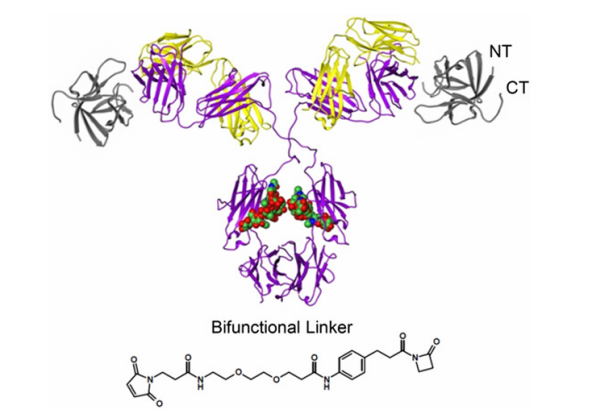
 

Structure and design mechanism of PF-05231023 [1]

PF-05231023 (AbMole, M10048) significantly inhibits hepatic lipid accumulation in animal models, reduces adipocyte hypertrophy in white adipose tissue, and ameliorates lipid metabolism disorders by downregulating lipid synthesis-related genes in adipose tissue transcriptomes[4]. Moreover, PF-05231023 reduces body weight in animal models by affecting the central nervous system and decreasing food intake. Studies have confirmed that the weight loss induced by PF-05231023 in cynomolgus monkeys and diet-induced obese mice is primarily due to reduced food consumption[5].

PF-05231023 is also used in animal models of nonalcoholic steatohepatitis (NASH). In diet-induced NASH mouse models, PF-05231023 significantly reduces body weight, hepatomegaly, plasma transaminase levels, and plasma/hepatic lipid levels, while also improving liver fibrosis [6]. The mechanism involves inhibition of hepatic lipid droplet accumulation and reduction of adipocyte hypertrophy in white adipose tissue.

PF-05231023 has important applications in animal models of diabetes and its complications. For example, in insulin-deficient diabetic mice, PF-05231023 activates the AKT-Nrf2 pathway, reduces IL-1β expression in photoreceptor cells, protects retinal neuron function, and ameliorates diabetes-induced retinal dysfunction and photoreceptor segment disruption [7].


In 2014, two inhibitors from AbMole were used in in vivo studies by the Spanish National Center for Cardiovascular Research (CNIC) and Columbia University, leading to research publications in Nature and Nature Medicine.


Case Study

J Adv Res. 2025 Mar;69:477-494.

A research team from Wenzhou Medical University investigated the role and mechanism of FGF21 in a mouse model of septic liver injury. They found that FGF21 inhibits septic liver injury via the autophagy/HIF-1α axis. Specifically, FGF21 activates the FGFR receptor, inhibits the mTOR signaling pathway, restores impaired autophagic flux in macrophages during sepsis, promotes p62-mediated autophagic degradation of HIF-1α, and reduces pro-inflammatory cytokine release and hepatic inflammatory infiltration. PF-05231023 (AbMole, M10048), a long-acting FGF21 analog, was used to inhibit pathological progression and significantly improve survival in septic mouse models. Furthermore, PF-05231023 effectively inhibited the activation of pro-inflammatory macrophages in mice [8].
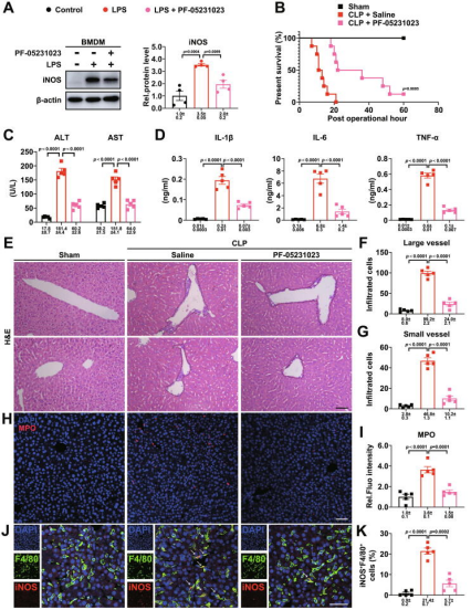
 

PF-05231023 protects mice against septic liver injury [8]

The products described herein are for research use only.


References and Acknowledgments

[1] Jie Huang, Tetsuya Ishino, Gang Chen, et al., Development of a novel long-acting antidiabetic FGF21 mimetic by targeted conjugation to a scaffold antibody, 346(2) (2013) 270-280.

[2] Junming Yie, Randy Hecht, Jennifer Patel, et al., FGF21 N-and C-termini play different roles in receptor interaction and activation, 583(1) (2009) 19-24.

[3] J. Huang, T. Ishino, G. Chen, et al., Development of a novel long-acting antidiabetic FGF21 mimetic by targeted conjugation to a scaffold antibody, The Journal of pharmacology and experimental therapeutics 346(2) (2013) 270-80.

[4] J. Zhao, X. Liu, J. Yue, et al., PF-05231023 reduces lipid deposition in apolipoprotein E-deficient mice by inhibiting the expression of lipid synthesis genes, Frontiers in veterinary science 11 (2024) 1429639.

[5] W. C. Thompson, Y. Zhou, S. Talukdar, et al., PF-05231023, a long-acting FGF21 analogue, decreases body weight by reduction of food intake in non-human primates, Journal of pharmacokinetics and pharmacodynamics 43(4) (2016) 411-25.

[6] M. H. Nielsen, M. P. Gillum, N. Vrang, et al., Hepatoprotective effects of the long-acting fibroblast growth factor 21 analog PF-05231023 in the GAN diet-induced obese and biopsy-confirmed mouse model of nonalcoholic steatohepatitis, American journal of physiology. Gastrointestinal and liver physiology 324(5) (2023) G378-g388.

[7] Z. Fu, Z. Wang, C. H. Liu, et al., Fibroblast Growth Factor 21 Protects Photoreceptor Function in Type 1 Diabetic Mice, Diabetes 67(5) (2018) 974-985.

[8] J. Zhu, Z. Jin, J. Wang, et al., FGF21 ameliorates septic liver injury by restraining proinflammatory macrophages activation through the autophagy/HIF-1α axis, Journal of advanced research 69 (2025) 477-494.


AbMole Mini-Lecture丨Pepstatin A: An Aspartic Protease Inhibitor and Its Multifaceted Research Applications


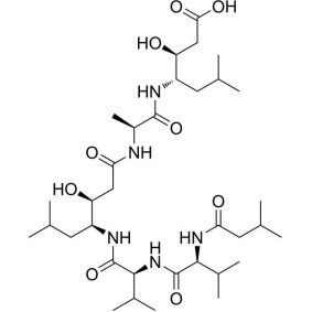
Pepstatin A (AbMole, M3657) is a naturally occurring peptide compound produced by Actinomycetes. Its chemical structure features two unique statine residues ((3S,4S)-4-amino-3-hydroxy-6-methylheptanoic acid), which enable potent inhibition of aspartic protease family members. X-ray crystallography studies have shown that the hydroxyl group of the statine residue in Pepstatin A (CAS No.: 26305-03-3) forms a stable hydrogen bond network with the two catalytic aspartic acid residues in the active site of aspartic proteases (with an inhibition constant Ki as low as 0.072 μM). This mode of interaction endows Pepstatin A with the ability to inhibit enzymes such as pepsin, cathepsin D, and cathepsin E[1].

AbMole provides global researchers with high-purity, high-bioactivity inhibitors, cytokines, human monoclonal antibodies, natural products, fluorescent dyes, peptides, compound libraries, antibiotics, and other research reagents, widely cited in numerous publications and patents worldwide.


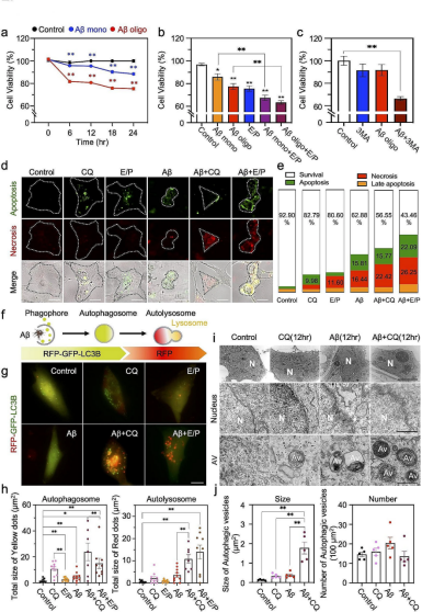
In experimental settings, Pepstatin A (AbMole, M3657) is frequently used in autophagy-related research, often in combination with E-64d (Aloxistatin, AbMole, M2376) to block the lysosomal degradation pathway. This inhibitor combination effectively blocks the final degradation step of the autophagic flux by inhibiting lysosomal proteases, leading to the accumulation of autophagosomes and lysosomes within cells. For example, co-treatment with Pepstatin A and E-64d induced the accumulation of the autophagy marker LC3-II in starved HEK293 and HeLa cells, indicating blockade of autophagic flux [2]. Pepstatin A also exhibits diverse inhibitory activities in viral replication suppression. Studies have demonstrated that Pepstatin A effectively inhibits the activity of viral proteases (e.g., ASPRV1 protease), thereby restricting viral replication[3]; it also interferes with viral hydrolysis of chemokines by inhibiting cathepsin D activity, affecting HIV replication [4]; furthermore, Pepstatin A broadly suppresses viral replication by upregulating apoptosis-associated tyrosine kinase (AATK), and AATK knockout significantly attenuates the antiviral effect of Pepstatin A[5]. Pepstatin A also influences amyloid precursor protein processing via inhibition of cathepsin D. For instance, in APP/PS1 transgenic mouse models, Pepstatin A (10 mg/kg) reduced β-amyloid plaques in the cerebral cortex by 35%, accompanied by downregulation of the microglial activation marker CD68[6], offering new perspectives for mechanistic studies in neurodegenerative animal models such as Alzheimer’s disease. Pepstatin A also induces inhibition of RANKL-induced osteoclast differentiation in a dose-dependent manner, an effect that is independent of cathepsin D and other proteases and primarily involves blockade of the ERK signaling pathway [7].

In 2014, two inhibitors from AbMole were used in in vivo studies by the Spanish National Center for Cardiovascular Research (CNIC) and Columbia University, leading to research publications in Nature and Nature Medicine.


Blockage of autophagy pathway decreases survival of astrocytes in response to Aβ oligomer [6]

The products described herein are for research use only.


References and Acknowledgments

[1] R. E. da Silva-Lopez, T. A. A. de Araujo, H. J. J. Monteiro, et al., Study of protease activity from Aspergillus awamori INCQS2B.361U2/1 extracellular fraction and modification of culture medium composition to isolate a novel aspartic protease, Brazilian journal of microbiology : [publication of the Brazilian Society for Microbiology] 53(3) (2022) 1599-1611.

[2] Isei Tanida, Naoko Minematsu-Ikeguchi, Takashi Ueno, et al., Lysosomal turnover, but not a cellular level, of endogenous LC3 is a marker for autophagy, 1(2) (2005) 84-91.

[3] M. Golda, J. A. Motyan, K. Nagy, et al., Biochemical Characterization of Human Retroviral-Like Aspartic Protease 1 (ASPRV1), Biomolecules 10(7) (2020).

[4] O. A. Arodola, M. E. Soliman, Hybrid 2D/3D-quantitative structure-activity relationship and modeling studies perspectives of pepstatin A analogs as cathepsin D inhibitors, Future medicinal chemistry 10(1) (2018) 5-26.

[5] L. Zhao, R. Song, Y. Liu, Glycolytic metabolite phosphoenolpyruvate protects host from viral infection through promoting AATK expression, European journal of immunology 53(12) (2023) e2350536.

[6] Suhyun Kim, Heejung Chun, Yunha Kim, et al., Astrocytic autophagy plasticity modulates Aβ clearance and cognitive function in Alzheimer’s disease, 19(1) (2024) 55.

[7] Hajime Yoshida, Kuniaki Okamoto, Tsutomu Iwamoto, et al., Pepstatin A, an aspartic proteinase inhibitor, suppresses RANKL-induced osteoclast differentiation, 139(3) (2006) 583-590.


AbMole Mini-Lecture | Lauroyl-L-carnitine: A Metabolite with Multiple Roles as a Signaling Molecule and Compound Carrier


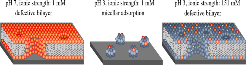
Lauroyl-L-carnitine (AbMole, M58111) is a medium-to-long-chain acylcarnitine that plays complex and critical roles in cellular metabolism and signal transduction. Metabolically, Lauroyl-L-carnitine serves as an activated carrier of lauric acid (C12 fatty acid), shuttling lauroyl groups from the cytoplasm across the inner mitochondrial membrane into the mitochondrial matrix for β-oxidation to fuel cellular activities[1]. This mechanism is fundamental for maintaining cellular energy homeostasis. When fatty acid oxidation is impaired or this molecule abnormally accumulates, it transitions from a simple metabolic intermediate into a biologically active signaling molecule. Studies show that Lauroyl-L-carnitine’s biological activity is primarily mediated through modulation of specific ion channels. It is an agonist of the transient receptor potential ankyrin 1 (TRPA1) channel, whose activation is linked to calcium influx and subsequent calmodulin-mediated pathways [2]. Additionally, abnormally elevated Lauroyl-L-carnitine induces mitochondrial dysfunction [2]. Lauroyl-L-carnitine has also been confirmed as a ligand for the G protein-coupled receptor GPR84, involved in regulating inflammation-related signaling pathways[3]. In experimental design, Lauroyl-L-carnitine serves as an important tool for probing metabolic networks and cellular stress responses. On the other hand, in animal studies, Lauroyl-L-carnitine reduces solution surface tension, enhancing the dissolution and diffusion of compounds and bioactive molecules in the gastrointestinal tract, thereby promoting their absorption. The mechanism is twofold: first, its amphiphilic structure (hydrophobic lauroyl chain + hydrophilic carnitine head group) allows it to form micelles in aqueous solutions and encapsulate hydrophobic bioactive molecules; second, Lauroyl-L-carnitine binds to the carnitine transporter OCTN2 on intestinal epithelial cells, facilitating targeted absorption of nanoparticles [4]. Compared with synthetic surfactants, Lauroyl-L-carnitine, as an endogenous metabolite, offers better biosafety. In 2014, two inhibitors from AbMole were used in in vivo studies by the Spanish National Center for Cardiovascular Research (CNIC) and Columbia University, leading to publications in Nature and Nature Medicine.


Figure 4. Micelle formation of Lauroyl-L-carnitine in buffers at different pH [5]


References and Acknowledgments

[1] M. Sisi, L. Yong, Q. Lixing, et al., L-carnitine: new perspectives on the management of preterm infants, Frontiers in nutrition 12 (2025) 1508441.

[2] H. Turk, S. Erdal, R. Dumlupinar, Exogenous carnitine application augments transport of fatty acids into mitochondria and stimulates mitochondrial respiration in maize seedlings grown under normal and cold conditions, Cryobiology 91 (2019) 97-103.

[3] H. Liu, X. Hu, Z. Li, et al., Surface adsorption and solution aggregation of a novel lauroyl-l-carnitine surfactant, Journal of colloid and interface science 591 (2021) 106-114.

[4] L. Kou, R. Sun, S. Xiao, et al., OCTN2-targeted nanoparticles for oral delivery of paclitaxel: differential impact of the polyethylene glycol linker size on drug delivery in vitro, in situ, and in vivo, Drug delivery 27(1) (2020) 170-179.

[5] Huayang Liu, Ke Fa, Xuzhi Hu, et al., Structural features of interfacially adsorbed acyl-l-carnitines, 623 (2022) 368-377.