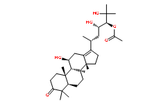
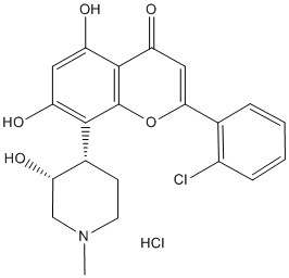
In addition, we provide genetic evidence that the molecular target of cocaine is the C. elegans SERT mod-5. These results also suggest that the observed cocaine response is not due to a non-specific local anesthetic effect of cocaine, which primarily results from its blockade of voltage-gated sodium channels. Locomotion is probably not the only worm behavior that can be modulated by cocaine. In C. elegans, serotonin regulates a wide variety of behaviors, including egg-laying, feeding, chemosensation, male turning, and learning and memory. Thus, it remains possible that cocaine may also modulate other types of worm behaviors. In rodents, cocaine can target all major types of monoamine transporters, including DAT, SERT and NET. Surprisingly, we did not detect a significant role for dopamine in cocaineinduced locomotor response, considering that acute dopamine treatment has also been demonstrated to inhibit worm locomotion. Nevertheless, it remains possible that dopamine may play a role in mediating cocaine response in C. elegans but such a role is not manifested in our assay. The response to cocaine in C. elegans requires the ionotropic serotonin receptor MOD-1, suggesting MOD-1 as a downstream effector of cocaine. Since MOD-1 is an inhibitory Cl2 channel, this suggests that the cocaine-induced hypolocomotor response may result from MOD-1-mediated Acotiamide hydrochloride inhibition of locomotion. Indeed, MOD-1 has been shown to mediate serotonin-induced paralysis of C. elegans. In rodents, one of the major downstream targets of cocaine is the 5-HT1A-receptor, which couples via Gi/Go to a hyperpolarizing K + conductance, and is thus inhibitory. Therefore, in both worms and mammals cocaine appears to evoke a serotonin-mediated response through inhibition of neurotransmission. Our findings shed light on questions surrounding the involvement of serotonin in mediating the behavioral effects of psychostimulant drugs such as cocaine. A growing body of evidence demonstrates that in addition to dopamine, serotonin plays an important role in mediating behavioral and addictive effects of cocaine. Our results from C. elegans also support a critical role for serotonin in cocaine responses. Although at the behavioral level cocaine elicits distinct responses in worms and mammals, at the molecular level this drug impinges on similar types of genes and pathways in both organisms, suggesting that C. elegans may be used to study the mechanisms by which serotoninergic signaling regulates cocaine responses. The most common disease of the AV is calcific aortic stenosis, found in 2% of individuals over 65 years and in 4% of those over 85. Early lesions with some features of atherosclerosis are NGP 555 found in almost all adults.These lesions may progress into calcified nodules, which can grow over time, stiffening the valve leaflets and eventually critically interfering with valve opening and potentially closing. Currently, the most common treatment for CAS is valve replacement with a mechanical or bioprosthetic valve. CAS is the leading single etiology of valve disease necessitating replacement, accounting for a major fraction of the approximately 300,000 valve replacement surgeries worldwide each year. Overall valve function depends on the mechanical properties of the cuspal tissue: stiffer, thicker tissue causes the valve to be less efficient. A model that describes the connection between tissue properties and valve function will be clinically useful in two ways.
Monthly Archives: July 2019
Non invasive imaging methods using protease-specific contrasts agents are natural candidates for this purpose
From the point-of-view of integrative biology, the question arises whether those events could be detected and followed in-vivo in an intact organism by revealing the corresponding proteolytic activity. Furthermore detecting an enzymatic activity offers the possibility of signal amplification via a renewable substrate. The first evidence that protease imaging is a pertinent way to study diseases in vivo was given using near infrared fluorimetry on a murine tumor model. Self quenched fluorescent peptides were actually cleaved and were able to generate a detectable signal in the tumor environment. In spite of recent progresses optical methods have strong limitations due to the light transmission to and from deep-seated organs. With this respect Magnetic Resonance Imaging constitutes a good alternative. Interesting IPI3063 protease and glycosidase substrates acting as MRI contrast agents have been proposed. However lower toxicity and much higher contrasts are needed to compensate for the low sensitivity of nuclear magnetic resonance. Overhauser Magnetic Resonance Imaging has the potential to significantly enhance the sensitivity of MRI. It is a double resonance experiment that transfers a fraction of the higher magnetization of the electron of a free radical to the protons of surrounding water molecules. Recently OMRI was successfully applied to in vivo oxymetry imaging by correlating the Electron Paramagnetic Resonance line width variation of a trityl free radical to oxygen concentration. Nitroxides are a family of stable free radicals. Several biocompatible nitroxides have been used in EPR and OMRI experiments in vivo. The Overhauser enhancement strictly depends on the nitroxide EPR line width. Due to the nitroxide asymmetric structure their EPR spectra significantly widen and flatten as their rotational correlation times increase. Here this property was applied to design a contrast agent sensitive to proteolysis and detectable through Overhauser enhancement. In this paper a general molecular imaging method with generation of high positive contrast in the presence of proteolytic activity is proposed. As an example nitroxides were covalently bound to bovine serum albumin. An experimental setup for OMRI at 0.2 T was built so that dynamic nuclear polarization can occur at depth in the range of a centimeter without any significant heating of the sample. OMRI of the nitroxide-labeled BSA sample PF-06651600 revealed no signal enhancement because of the elevation of the nitroxide correlation time. The initial magnetic resonance image intensity was strongly enhanced by enzymatic digestion of the carrier protein. Such a protease-switch method can be adapted to any proteolytic activity by linking a nitroxide to a large carrier molecule through specifically cleavable peptide substrates. In this paper a non invasive method designed to perform MRI of the proteolytic activity in deep-seated organs is described. This method needs a magnetic resonance imaging system including a resonant cavity tuned on the free electron EPR frequency of a nitroxide. With a very simple biochemical model, it is demonstrated that a proteolytic enzyme can modulate the Overhauser effect through the alteration of the motional correlation time of a nitroxide labelled substrate.
The M1 muscarinic receptor antagonist and the M4 antagonist stricter FWE correction was used in this situation
In contrast, the effective size of the genotypic differences between healthy adults is expected to be small. Therefore, we used a relatively loose threshold to avoid missing the subtle differences between groups.  However, the intergroup differences cannot survive after the FWE correction for multiple comparisons. The lack of significant intergroup differences after a stricter FWE correction suggests that these findings should be validated in future studies. In summary, this study used a relatively large sample size of healthy young adults and a whole brain analyzing method. We found that the COMT Val158Met polymorphism modulates anatomical morphology and related rsFCs within the DMN, indicating a potential neural pathway by which this polymorphism may affect cognitive function. Meanwhile, we found a genotype �� gender interaction in the prefrontal GMV but not in the GMV of the PCC and the rsFCs within the DMN. The mechanisms of these findings need to be further investigated. Fragile X syndrome is the most common inherited cause of intellectual disability and the most common singlegene defect identified in autism. Approximately one-third of patients with FXS are eventually diagnosed with autism spectrum disorder, and it is estimated that up to 6-8% of children diagnosed with autism carry mutations in the X-linked FMR1 gene. Patients with FXS often have deficits in verbal and performance skills, spatial reasoning, and short term memory, as well as attention deficits and hyperactivity, stereotypic movements, and atypical social development. FXS results from inappropriate transcriptional silencing of the FMR1 gene and failure to express its product, FMRP, an RNA-binding protein that represses local protein synthesis. Mice lacking the Fmr1 gene model aspects of the pathophysiology and many of the abnormal behaviors seen in FXS and autism, including cognitive impairments, increased spontaneous motor activity, increased seizure susceptibility; and altered social behaviors. FMRP opposes signaling through G protein-coupled Talazoparib company receptors acting through the Gq��-subunit, including group I metabotropic glutamate and M1 muscarinic acetylcholine receptors. Gq-coupled GPCRs signal through phospholipase-C and phosphoinositide 3-kinase, influencing local protein synthesis through both the Akt/ mTOR and MEK/ERK pathways. In dendritic spines, activity at these receptors in response to stimuli facilitates local synaptic protein translation; and lack of FMRP therefore leads to abnormally exaggerated experience and protein synthesis-dependent synaptic plasticity. Spine development is impaired such that spines are longer and thinner, retaining a more immature form, and do not undergo normal experience-dependent modification of size, shape, or number. PLC signaling is important in activitydependent spine development, supporting findings that mGluR antagonists normalize spine morphology in fmr1-null mice. Heterodimeric D1/D2 dopamine receptors also activate PLC through Gq, but whether this signaling mechanism is affected in Fmr1-null mice is not yet known. Although the pathophysiological mechanisms in FXS are some of the most understood among the genetic synaptopathies, MDV3100 therapy for this disorder currently consists of symptom management and not pharmacological correction or reversal of synaptic changes due to loss of FMRP. Both mAChR and mGluR-dependent LTD is enhanced in hippocampal neurons. Antagonism of mGluRs has been proposed as a rational therapy for FXS, and preclinical studies have shown that mGluR5 antagonists can partially correct some abnormal behaviors in Fmr1-null mice, including increased open-field exploration, impaired rotarod performance, and decreased prepulse inhibition, although results have been mixed. While still under clinical development, no mGluR antagonists are yet approved for human use.
Suggest a possible role of the Bmal2 gene in phase-shifting the hepatic clock in response to RF in SHR
For the other E-box driven genes studied herein, namely those involved in interactions between the clock and metabolism, i.e., Dbp, E4bp4 and Nampt, the advance was subtle and the amplitude was only slightly decreased. Similarly to that of Wee1, the expression profile of none of these genes did differ between the two rat strains under ad libitum feeding. Under RF conditions, the expression of Prkab2 exhibited a circadian oscillation in the SHR but was not rhythmic in the Wistar rats. The results also demonstrated that under RF, expression profiles of some studied genes in the liver of SHR were suppressed or did not differ compared with those in Wistar rats. This might be because these genes are not driven by the clock but rather by changes in the metabolic state. The RF likely affected the metabolic state of the SHR Perifosine moa differently than the Wistar rats because the constitutive expression of Pgc1��, which coordinates the transcriptional programs important for energy metabolism and homeostasis, was significantly suppressed in the SHR compared with the Wistar rats. Pgc1�� encodes a Temozolomide Autophagy inhibitor co-activator that enhances the activity of many nuclear receptors, including PPAR��. It is noteworthy that the Ppara expression was also slightly suppressed in the SHRs but became expressed rhythmically in response to RF, though with a low amplitude. The observed suppression was not due to a lower strain-specific basal expression of these genes under ad libitum conditions. The significant suppression of Pgc1�� and Ppara expression may likely result in decreased levels of the PPAR��/PGC1�� transcriptional  complex with nuclear receptors, which may consequently affect the activity of downstream genes regulating fatty acid uptake and oxidation in the liver of SHR maintained under RF. The other metabolism-relevant gene expression profiles in the liver did not differ between the two strains, namely that of Pparg, Hdac3, Hif1a and Ppp1r3c. Thus, the data suggest that RF impacts the metabolic state in the liver of SHR differently than in Wistar rats, with the most obvious difference in the suppression of Pgc1�� and Ppara expression and the imposition of a low-amplitude rhythmicity in Ppara and Prkab2 expression. These changes in gene expression might have a broad impact on the transcription of variety of genes, including those whose products feed back on the circadian clock mechanism. We hypothesize that as a consequence, apart from other effects, the transcriptional activity of the Bmal2 gene may also have changed. Based on the limited data available, the likely pathways for this effect are highly speculative. To test the hypothesis that the abnormal metabolic state of SHR under RF may account for the higher sensitivity of their hepatic circadian clock to changes in the feeding regime, future studies should examine the suggested pathways at the level of protein expression and activities. It is also reasonable to consider the involvement of glucocorticoids, which are known to play a role in resetting the phase of the peripheral clock in the liver and whose peak levels are shifted due to RF in rats. The general hypersensitivity of the hypothalamic-pituitaryadrenal system of SHR might theoretically account for the facilitation of the hepatic clock response to RF. However, similar to the previously suggested pathways, the role of glucocorticoids in this effect has not been tested. In conclusion, our results demonstrate a previously unknown correlation between the extent of FAA and the phase-shifting of the peripheral clock in the liver. Additionally, our evidence that the putative food-entrainable oscillator driving the FAA is more sensitive in SHR may facilitate future studies aimed at localizing this clock in the mammalian body.
Our previous work highlights the AhR as a key anti-inflammatory protein by an unknown mechanism
However, the AhR profoundly controls the nuclear levels of HuR in response to CSE. HuR translocation from the nucleus to the cytoplasm is critical to its ability to stabilize target mRNA. This may be why in AhR+/+ cells, where HuR remains within the nucleus, HuR knockdown had no effect on Cox-2 mRNA stability. Results in C5N cells, a mouse  keratinocyte cell line with exclusive nuclear HuR support this notion, as reduction in HuR expression had no effect on ornithine decarboxylase mRNA stability. However in AhR2/2 cells whereby HuR translocates to the cytoplasm, HuR was a key factor involved in Cox-2 mRNA stability, as siRNA-knockdown resulted in enhanced Cox-2 mRNA degradation. Our results support that retention of nuclear HuR is an important feature in the destabilization of Cox-2 mRNA by the AhR. In addition to Cox-2, HuR has thousands of target genes and stabilizes mRNAs that encode proteins associated with a variety of cellular functions including cell cycle, proliferation, apoptosis and inflammation. The AhR regulation of these functions is established opening the possibility that AhR retention of nuclear HuR may have important implications for the regulation of genes beyond the control of Cox-2. Our results are also the first to show in vivo evidence of pulmonary HuR translocation in response to cigarette smoke. In the lungs of AhR2/2 mice, there was no Cox2 mRNA induction despite concordant COX-2 protein and profound cytoplasmic HuR. It was surprising to note considerable levels of HuR in the cytoplasm of pulmonary cells without smoke exposure. Cytoplasmic HuR has been reported in the lungs of adult A/J mice, consistent with our data, and HuR expression is required for proper lung development. It may be that in the lung, an organ continuously exposed to the environment and one that is highly susceptible to Tubacin oxidative damage, a constitutive level of cytoplasmic HuR is required to ensure optimum immunological responsiveness. Although our results reveal a novel pathway in which the AhR regulates COX-2 protein expression by controlling the cellular localization of HuR, it remains to be established precisely how the AhR retains HuR in the nucleus. Our finding that the AhR regulates HuR localization in response to CSE, but not BP indicates divergent mechanism of AhR activation in maintaining HuR localization despite the ability of both to cause Cyp1a1 mRNA induction. It also indicates that BP, which is present in cigarette smoke, is not the component causing HuR translocation to the cytoplasm in the absence of AhR expression. Cigarette smoke is a complex mixture, containing more than 4800 compounds including metals, oxidants and free radicals, the latter of which are a potent source of oxidative stress. Given that the AhR protects against oxidative damage due to smoke exposure, it reasonable to speculate that the high oxidant conditions exerted by cigarette smoke contributes to HuR translocation in the absence of AhR expression. AhR activity was required to retain HuR within the nucleus, but did not require DNA-binding. It has been speculated that the DREindependent anti-inflammatory abilities of the AhR may involve multiple protein-protein interactions. The AhR localizes to the nucleus in the absence of exogenous ligand, a cellular phenomenon that depends on cell-cell contact. Adherent cells grown to sub-confluence, methodologically similar to the experiments conducted here, exhibit both cytoplasmic and nuclear AhR, making interaction with AhR and HuR within the nucleus a plausible assumption. Thus, while there is no known physical association between AhR and HuR, it is interesting to speculate that the AhR may interact with HuR to prevent its nuclear export, a notion we are actively Everolimus pursuing. It is believed that the AhR plays an important role in physiology independent of its ability to respond to dioxin.